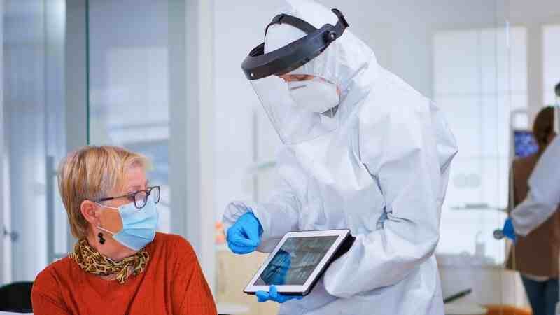
In medical imaging, especially in fields like dentistry, radiolucency vs radiopaque, and orthopedics, the terms radiolucency and radiopaque play a crucial role in diagnosis and treatment. These terms describe how different tissues or substances within the body interact with X-rays and other forms of medical imaging. Understanding the distinction between radiolucent and radiopaque materials is vital for professionals to correctly interpret diagnostic images.
What is Radiolucency?
Radiolucency refers to the property of a material that allows X-rays to pass through it relatively easily, making the area appear darker on the radiographic image. This is a key concept for professionals, as radiolucent materials are usually less dense, which is why they allow more radiation to pass through.
Common Radiolucent Materials
- Air: In a chest X-ray, the lungs appear dark because air is radiolucent.
- Fat and soft tissues: These materials have lower density, allowing some X-rays to pass through, but not as much as air. This results in a gray appearance on an X-ray.
- Fluid-filled cavities: Fluids like blood or cystic fluid are also radiolucent to a degree and often show as darker areas in the context of denser surrounding tissue.
Applications of Radiolucency in Medical Imaging
Radiolucency is vital for diagnosing various conditions:
- Lung pathologies: Radiolucency is essential for detecting abnormalities such as pneumothorax, where air escapes into the pleural cavity.
- Bone fractures: In some cases, early fractures might appear as radiolucent lines on X-rays.
- Dental care: In dental X-rays, areas of decay show up as radiolucent regions due to the breakdown of tooth structure.
What is Radiopaque?
On the other hand, radiopaque substances are those that do not easily allow X-rays to pass through, meaning they appear lighter or white on the radiographic image. These materials are dense, creating a more significant barrier to radiation.
Common Radiopaque Materials
- Bone: One of the most common radiopaque materials, bones show up as bright white areas on X-rays.
- Metal implants: Surgical hardware such as screws, plates, or dental fillings is highly radiopaque.
- Calcium deposits: These can be seen in the form of calcified tissues, such as kidney stones or atherosclerotic plaques, that block X-rays effectively.
- Contrast agents: In some medical procedures, contrast agents like barium or iodine are used to enhance imaging, as they are highly radiopaque and outline structures clearly.
Applications of Radiopaque in Medical Imaging
Radiopaque materials are essential in visualizing:
- Bone structure: Radiopaque bones are key in diagnosing fractures, bone malformations, and monitoring healing after surgical interventions.
- Dental restorations: Dental X-rays frequently show radiopaque fillings or crowns, helping dentists evaluate the success of dental treatments.
- Contrast studies: Radiopaque contrast agents enhance visibility of organs like the intestines, bladder, or blood vessels during specialized imaging procedures, such as barium swallows or angiograms.
Key Differences Between Radiolucency vs Radiopaque
Understanding the difference between radiolucent and radiopaque substances is crucial for accurate interpretation of medical images.
- Appearance on X-rays:
- Radiolucent: Dark or black areas, representing substances that X-rays can pass through easily.
- Radiopaque: White or light areas, indicating denser substances that block X-rays.
- Physical Composition:
- Radiolucent materials: Air, fluids, and soft tissues.
- Radiopaque materials: Bone, metal, and calcifications.
- Diagnostic Implications:
- Radiolucent areas may indicate problems such as tissue loss, fractures, or cysts, whereas radiopaque areas may signal bone structures, foreign objects, or calcifications.
How Medical Professionals Use Radiolucency and Radiopaque Properties
Medical professionals use the contrasting properties of radiolucency and radiopaque in several ways to enhance diagnosis and treatment:
- Dental Care: In a dental exam, radiopaque fillings and bone structure allow for the detection of cavities or fractures in radiolucent areas.
- Orthopedics: X-rays of bones and joints highlight radiopaque bones, allowing doctors to assess fractures, deformities, or foreign objects.
- Oncology: Radiolucent tumors may show up as dark masses within denser radiopaque tissues, aiding in early cancer detection.
- Trauma Assessment: Radiolucency can reveal gas accumulation in tissues (e.g., pneumothorax), while radiopaque substances like foreign objects (e.g., bullets, shrapnel) become easily identifiable.
Common Misinterpretations in Radiography
Misinterpretations between radiolucent and radiopaque areas can lead to diagnostic errors. For instance, a small radiolucent lesion in a bone might be missed or mistaken for a benign feature if not properly examined. Conversely, a calcified, radiopaque deposit might be misdiagnosed if its clinical significance is overlooked.
Importance of Training and Expertise
A radiologist’s ability to distinguish between radiolucent and radiopaque materials directly impacts patient outcomes. Continuous training, along with the use of advanced imaging technologies, can significantly reduce misinterpretation rates in clinical practice.
Advancements in Imaging Techniques
Modern imaging techniques, such as CT scans, MRI, and ultrasound, complement traditional X-rays by offering more detailed views of both radiolucent and radiopaque structures. For instance:
- CT scans provide cross-sectional images that clearly differentiate between soft tissue, bone, and foreign objects.
- MRI is excellent for imaging soft tissues that may not be visible on traditional X-rays.
- Ultrasound uses sound waves, which can provide additional context for evaluating radiolucent areas filled with fluid or soft tissue abnormalities.
Conclusion
In the field of medical imaging, understanding the terms radiolucency vs radiopaque is fundamental for accurate diagnosis and treatment. By leveraging the contrasting properties of different tissues and materials, medical professionals can visualize internal structures more effectively. Whether you’re a radiologist, dentist, or orthopedic surgeon, recognizing the significance of these terms is crucial for successful patient outcomes.

